Stroke Rehabilitation
Stroke rehabilitation is the specific process of rehabilitating someone who has suffered a stroke. The basic ideas of neuroplasticity and how recovery occurs in the brain are discussed in this chapter to show how motor control and learning concepts may be applied. The chapter will include the present state of stroke rehabilitation, the sorts of therapies utilised, and, lastly, mirror therapy, which is the main therapy that the system is designed around. Stroke rehabilitation integrates elements identified in chapter 1 into a rehabilitation programme for the patient to do to successfully recover from the stroke.
Neuroplasticity
Human brains are malleable due to plasticity and can adapt to the environment. Neuroplasticity is the mechanism that allows stroke patients to be able to recover from impairments. With regards to the distribution of motor cortex volume, “The representation of the body is distorted, with a disproportionate volume of cortex devoted to the hand” [42]. There are two important points to draw from this. Firstly, the hands are for human motor skills and secondly, since there is a disproportionate allocation of volume, if there is an injury to the cortex it will be more likely hand function will be impacted in comparison to other body parts. Also, a lot of rehabilitation is task orientated. These six principles to optimise training are: specificity of training; constrained use of the impaired limbs; mass practice (repetition); shaping of skill; saliency of task; knowledge of performance and results [43].
Neuroplasticity presents an opportunity to take advantage of that mechanism by activating mirror neurons and reinforcing recently learned movements to aid in rehabilitation and recovery. By tracking brain activity in specific areas, through such means as transcranial magnetic stimulation (TMS), it is possible to monitor any changes that take place for adaptation and to witness the reorganisation of connections to responses. TMS is a non-invasive procedure, which uses the magnetism. Recent neuroimaging and TMS studies show that the primary motor area (M1) was activated by the observation of movement and imagery of movement [44]. The early presence of the Motor Evoked Potential (MEP) for predicting a “good prognosis is generally accepted, and motor recovery is associated with improved corti-cospinal conduction.” [45]. MEP are electrical signals recorded from the motor pathways. When recording the MEP data, caution must be taken when measuring the most appropriate pathway, as it could be difficult to get the correct readings [46]. For stroke sufferers, neuroplasticity plays a major role in rehabilitation to help undo the damage caused to sensory and motor function. Murphy and Corbett’s study [47] provides evidence to suggest that Hebbian processes play a critical role in creating activity-dependent changes in synaptic strength. Direct evidence for Hebbian processes after stroke is sparse, just as it is for homeostatic mechanisms.
There are many complex mechanisms involved in regrowth and regeneration of cells. This is referred to as neurogenesis, comprising several steps, occurs in the hippocampus and subventricular zone. Mossy-fibre sprouting, axonal sprouting in the peri-infarct region, dendrites and a gamut of neurochemicals and structures, all interact to permit new cell development, and reverse damage where possible and disinhibit areas previously inhibited [48]. Although younger brains appear to be more readily adaptable, older brains are also capable of significant reorganisation, which may occur quickly. Neurological mapping has demonstrated how much subjective qualia (individual instances of subjective, conscious experience) influence brain activity. Nummenmaa et al. [49] state that, “The intensity of the mental experiences and bodily sensations associated with subjective feeling states were highly correlated.” This information might help further research on neuropsychology and sensory psychology investigation and provide a greater understanding of the role conscious experience plays upon neural activity. It also offers another aspect for consideration in game design on how the patients might ‘feel’, which could assist their recovery. The lesions developed through stroke will determine the magnitude of their effect [50]. Small, multiple lesions are easier for the brain to deal with than a single large one. Whether the location is found in the anterior or posterior area of the brain will determine how motor or language function is affected, especially speech. The location of the original injury is significant in determining the potential symptoms associated with damage to a specific region, as well as how other neural pathways may be affected by damaged circuitry some distance away. An inability to receive signals could lead to malfunction of an earlier process dependent on these connections, atrophy or dendritic and synaptic deterioration through a reduction in density, or neuronal death resulting from apoptosis.
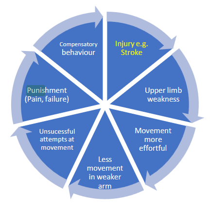
It is important to address the learned non-use (Fig.) [51] of the patient to make sure that they recover correctly. Learned non-use is defined as “Observed reduction of a behaviour that cannot be exclusively attributed to an impairment in the central nervous system”[6]. If they neglect movement, the limb will become weaker. A weaker limb requires more effort and as performance declines, the patient will mask the movement to compensate. This negative cycle will continue. Stress can impact recovery by affecting changes in brain plasticity, which has been shown to have significant consequences on the treatment of stress-related mental illness [52]. Treatment with CDP-choline, for example, enhances functional recovery and boosts neural plasticity after experimental stroke [53].
Repetitions are for the enforcing of neuroplasticity throughout the rehabilitation process, as it is not necessarily muscle weakness which is the issue but rather the signal pathway. If patients neglect training, their current state will deteriorate and learned non-use may occur. If we regularly have to perform a very skilled motor task, the cortical representation for the muscles involved will remain enlarged [54].
Motor Control and Learning
Understanding motor control and learning are essential to developing an appropriate system as the developer needs to understand what the purpose is, how it works and how to measure it. Misunderstanding this can lead to peculiar solutions, and although it works technically, it might not translate well into its intended task. For example, a Virtual Reality game where the patient throws a ball with a pinch motion gesture is not suitable as a throwing action, which involves the opening hand. A pinch motion is an unnatural movement when throwing. This conflict is not productive to the goal.
In consideration of how we move, learn to move, and relearn to move, it is important to understand the basic principles that facilitate motor performance. Continuous/serial/discrete movements [55] refer to the degree to which the movement has a distinct beginning and end. Each of these categories possesses a different level of difficulty relative to its execution, requiring the brain to process information appropriately. A movement may also require definition as open/closed, referring to the extent to which the environment is predictable. Here the brain must deploy the most effective cue anticipation and rely on memory for selection. Most motor control tasks fall into one or more categories. In measuring movement, experimenters apply the following: consideration to a measure’s objectivity (how closely two independent observers achieve the same score), its sensitivity to changes in skill requirements, its reliability (how well the score is repeatable), and its validity (how well the test measures its intended measure). Movements in terms of the environmental goal outcome use four basic measures in terms of: errors, speed, time or magnitude, or with various secondary tasks. According to Schmidt [55] the principal ways to measure errors in the movement are:
- Constant error (CE, a measure of average error or bias) 𝐶𝑜𝑛𝑠𝑡𝑎𝑛𝑡 𝐸𝑟𝑟𝑜𝑟 = 𝐶𝐸 = ∑( 𝑥𝑖 − 𝑇) / 𝑛
- Variable error (VEr, a measurement of inconsistency) 𝑉𝑎𝑟𝑖𝑎𝑏𝑙𝑒 𝐸𝑟𝑟𝑜𝑟 = 𝑉𝐸𝑟 = √∑(𝑥𝑖 −𝑀)2 /𝑛
- Total variability (E, a measure of overall error) 𝑇𝑜𝑡𝑎𝑙 𝑣𝑎𝑟𝑖𝑎𝑏𝑖𝑙𝑖𝑡𝑦 = 𝐸 = √∑(𝑥𝑖 −𝑇)2 /𝑛
- Absolute error (AE, also a measure of overall error). 𝐴𝑏𝑠𝑜𝑢𝑙𝑡𝑒 𝑒𝑟𝑟𝑜𝑟 = 𝐴𝐸 = ∑|𝑥𝑖 − 𝑇| / 𝑛
Different aspects of the measurement process utilise the error measures. Speed measures come into play when accuracy is less important (or is controlled) and when rapid actions are critical. Analysis of simultaneous secondary tasks provides a measure of the performer’s spare capacity after he/she has devoted attention to a primary task. These error measures can help with both system development and game design for how the patient’s ‘success’ might best be judged [55]. Note VE was used in the original reference by Schmidt [55] but has been modified to VEr to avoid confusion between the terms Variable Error and Virtual Environment.
Common methods for movement measurement include the calculation of kinematic variables (position, velocity, acceleration) and the recording of the electrical activity of the muscles Electromyography (EMG). Digital technology advances permit kinematics assessment, with precise measurements of sensory-motor control and studies of brain activity are no longer unusual. Using TMS to determine the force of the muscle used, an increase can be indicative of improved strength. The execution speed of a movement might indicate improved mobility, as would the ability to perform that task from different angles or other introduced variables that emulate activities of daily living [55]. An essential component of motor learning is feedback. Muscles, sensors involved in proprioception, and brain regions that respond to stimuli can all sense inherent feedback. Sometimes we are unaware of these feedback-related behaviours. Sensory information provides the bulk of feedback for movement, whether it is inherent or augmented. Inherent or intrinsic feedback is integral to tasks performed; it comes from within and may be visual, auditory, haptic, tactile, proprioceptive, balance, taste smells and goals and is often immediate. Augmented feedback also referred to as extraneous feedback falls into two classifications – knowledge of results (KR) and knowledge of performance (KP) – and these types of feedback naturally experience delays in their arrival [55]. When feedback occurs, it can be referred to as real time or terminal. Real time feedback is feedback which is happening at the same time as the activity. Terminal feedback is when the activity is completed.
Once the principles of motor control and learning are understood, then the designer of the system can appreciate the mechanisms and how it works. When this is known the designer can obtain a better understanding of what a therapist is trying to achieve with rehabilitation.
Traditional Stroke Rehabilitation
Rehabilitation is essential for stroke survivors to recover correctly. Post-stroke physical recovery requires muscle-strengthening and brain retraining to renew motor movement [56]. Rehabilitation achieved through intense, repetitive physical exercises targeted at motor recovery utilises brain neuroplasticity to reinforce relevant neural pathways [56]. Reduced motor function due to stroke affects mobility and limits daily life activities, participation in society, and diminishes the odds of returning to professional activities. All these factors contribute to the overall low quality of life and can contribute to frailty. Rehabilitation training is the most effective way to reduce motor impairments in stroke patients [57]. Technological solutions have been used for stroke rehabilitation which includes Virtual Reality (VR). Page and Peters [58] proposed the PRACTICE approach, which is a set of principles “based on converging findings from neuroscience and motor learning research” [58] to increase upper extremity function and acknowledge that many of the therapies investigated by researchers may be beneficial, but they come at a price which is stated [58].
“Few therapeutic approaches were showing promise in animal or early human trials fully translated to regular clinical practice. Consistently, many contemporary motor rehabilitation strategies require high duration contact time and costly equipment, preventing their widespread clinical application, except among specialized rehabilitation and academic medical centres.” Regarding upper limb interventions, stroke guidelines recommend a daily minimum of 45 minutes of therapy [59] over a minimum of 5 days a week [60]. Although post-stroke individuals have a rehabilitation programme provided to them, stroke survivors engage in less than four minutes during physiotherapy and eleven minutes during occupational therapy in acute rehabilitation[61]. Stroke survivors perform few activity-related arm movements [61], which is much lower than recommended. Even in a hospital, inactivity is a problem for stroke patients [62]. Often, patients are bed-bound and can only leave with supervision if they are deemed capable of ‘walking’ with assistance to the bathroom. Even for those with reasonable mobility, seldom do patients wander about the ward independently. They are either waiting for the doctor’s rounds, eating meals, conforming to their rest periods, or waiting for visitors – in their beds. Many stroke patients are inactive and alone while in hospital and as a consequence, it is imperative to improve activity levels and the effectiveness of interventions to increase physical activity after stroke [62]. Despite efforts to increase engagement with physiotherapy, the “Circuit class therapy and 7-dayweek therapy for Increasing Rehabilitation Intensity of Therapy after stroke trial” (CIRCIT) [63] discovered that inactivity between sessions was still a problem with patients. Participants were alone for almost half the working day, regardless of the model of physiotherapy.
Understanding how inactivity might harm recovery when physiotherapy sessions are viewed as the only physical activity in which patients participate. Kwakkel [64] suggests increasing the efforts of physiotherapy sessions and even incorporating off-duty practice during patient downtime. The more rigorous the practice following a stroke, the better [65]. This is supported by a 2012 study [66], which expresses that the motor function of the arm after a stroke may be improved by increasing the intensity of training. Occupational therapists, as well as physiotherapists, are receptive to the use of technology as a tool for rehabilitation, for several important and persuasive reasons listed by Langan et. al. [67] who mention that technology can be harnessed to advance standards in clinical practice. Objective provision of feedback through the technology can be exploited for personal encouragement with instant feedback. This lends itself to goal setting, self-monitoring, enhanced engagement through a sense of empowerment and an honest reflection of evaluation of progress. Any objective measurements can provide data for professionals to analyse and review.
Evidence of which interventions work, the underlying mechanisms, and whether they could improve with the use of technology, is vital for the project before developing the system. Teasell et al. [68], quantify the quality of evidence (See Appendix C) according to randomised trial size and the resulting desirable effects with A, B or C ratings and evaluate specific available therapies via a detailed grading system (See Appendix D) [69]. Quality of evidence in the systematic review documented the quality of evidence synthesised within the reviews based on criteria considered within the GRADE (Grading of Recommendations Assessment, Development and Evaluation) [69] Criteria include:
- Risk of bias due to flawed design or conduct of studies.
- Imprecision (e.g. when confidence intervals for treatment effect are wide).
- Inconsistency (e.g. when point estimates vary widely, I² is large).
- Indirectness (e.g. variations in participants, interventions, comparisons and outcomes).
-
Publication bias (may be explored with the use of funnel plots and classed as not suspected, suspected, strongly suspected or very strongly suspected)
- Definitions[69]:
- High quality: when further research is very unlikely to change our confidence in the estimate of effect.
- Moderate quality: when further research is likely to have an important impact on our confidence in the estimate of effect and may change the estimate.
- Low quality: when further research is very likely to have an important impact on our confidence in the estimate of effect and is likely to change the estimate.
- Very low quality: when we are very uncertain about the estimate.
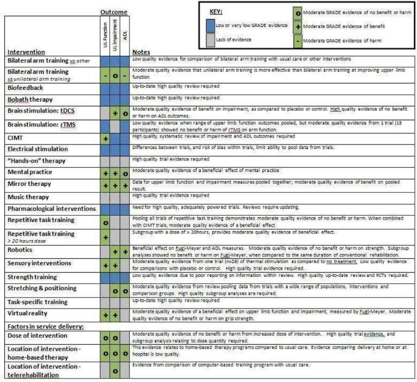
Key used for intervention outcome [69]:
- [Blue] Low or very low GRADE evidence
- [Grey] Lack of evidence
- [O] Moderate GRADE evidence of no benefit or harm
- [+] Moderate GRADE evidence of benefit
- [-] Moderate Grade evidence of harm
This research was conducted in 2019 during development. Fatigue is an important design consideration, and Eskes et al. [70] mention that “PSF (Post Stroke Fatigue) occurs frequently, has been associated with mood disorders and pain, and negatively impacts recovery after stroke”. Each stroke rehabilitee is unique, and this uniqueness puts demands on therapists to find the best fit for the individual under their care, which is important to remember, as it will also affect system design, making sure that program tailoring caters for the patient. Many rehabilitation treatment programmes are effective, which is why they were created. Lin and Dionn [71] state that functional performance improvement following a stroke does not favour one intervention over another. Rehabilitation specialists create tailored therapy regimens to maximise functional performance and involvement because stroke recovery is a dynamic process. When providing therapy services for the best patient outcomes, skilled therapy practitioners recognise the balance of art and science. Movement outcomes are influenced by several variables, such as stroke characteristics, intensity, social support, patient preferences, and goals.
In the UK, independent studies at UCL Queen Square [28] endorse much of the same evidence reviewed by the Canadian Stroke Best Practice [68] Recommendations team and suggest that much higher doses and intensity of upper limb neurorehabilitation can be delivered and should inform future clinical trial design. The study An Integrated Therapeutic Approach to Intensive Upper Limb Rehabilitation by K. Kelly and F. Brander [72] state the exact optimal dose required is not known. The best type of UL therapy is not known but likely each have their features. For motor learning the ‘learning’ requires the patient to learn how to do it. UL rehab cannot be simplified into a single protocol. More intensity appears better if it is low and the challenge is how to increase the intensity.
A review by Pollock et al. [69] covers 18 different types of interventions which included VR and Mirror Therapy (MT), as well as the dose of the intervention and the setting of intervention delivered, at the time of development this was the most recent review before the development of MINERVA and was used to review selected interventions. At present there is not high-quality grade evidence for any of the interventions currently used. Evidence of moderate quality indicates the potential effectiveness of interventions including VR and Mirror Therapy (MT). The only therapy that has shown a “Moderate grade of evidence of benefit” for outcome in all three areas (Upper Limb Function, Upper Limb Impairment and Activities of Daily Living (ADL)) is MT [69]. VR shows moderate grade evidence of benefit outcome for Upper Limb (UL) function and UL impairment. However, the benefits of ADL lack evidence (See (See Appendix D) for further details). Moderate-grade quality evidence also indicates that unilateral arm training may be more effective than bilateral arm training. Some evidence shows that a higher dose of an intervention is better than a lower dose. Additional research to identify the optimal dosage of arm rehabilitation is essential.
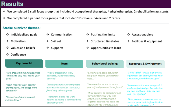
For an intervention to be successful, the dosage and a prescribed amount of work required for them to be able to recover are crucial. Increasing dosage beyond optimal can be detrimental as it could affect the recovery period, and as adherence is a problem, it may strain motivation to complete it. In specialist neurorehabilitation services patients may receive 5 hours of daily therapy, in others, 1 or 2 hours each day. The duration of therapy may vary from 2 weeks to 6 months [73]. Queen Square UL Programme [74] offers insight into the mechanisms of what could work well. Successful outcomes on modified upper limb Fugl-Meyer (FM-UL), which is a type of assessment used for motor recovery after stroke. These changes were maintained, or even improved upon, six months after treatment (Appendix E). Patients receive 100 hours of complete upper limb-focused therapy over the course of three weeks. Occupational and physical therapy sessions last two hours each day, with a particular emphasis on movement and task analysis, impairment treatment, and motor control re-education within functional tasks. Therapy is individualised and patients are urged to improve their cardiovascular fitness[74]. Currently, there are not enough programmes to know what is possible through motor/behavioural training. In summary, the dose of treatment is critical; more is deemed better. No evidence confidently claims which UL therapy is better at treatment, but likely each has its value. Simple task-specific training is not equivalent to learning and seems less useful. Motor Learning and re-education of motor control within functional tasks on how to complete it is required – with subjects who may lack capacity as well as capability. UL rehab cannot be reduced or simplified to a single protocol-based activity. The challenge is how to increase intensity across the neural pathway [74] as intensity appear related to outcome, “Clinically, greater intensity of stroke rehabilitation has been associated with improved outcome”[75]. The reflection of themes is summarised in Appendix F. It is worth noting the current expectations of treatment outcomes of task-specific training. Schoepp et al. studied the dose-response of task-specific upper-limb training in patients who had had a stroke for at least 6 months [76]. A finding was that “Overall, treatment effects were small. There was no evidence of a dose-response effect of task-specific training on functional capacity in people with long-standing upper limb paresis post-stroke.” Repetitive task training for improving functional ability after stroke BT - Cochrane Database Systematic Review [77] states: “Further research is needed to determine the best type of task practice and whether the more sustained practice could show better results.” To get an understanding of what is commonly done in most traditional rehabilitation. Connell et al. in “Prescribing upper limb exercises after stroke: A survey of current UK therapy practice,” [78] explain that low-intensity upper limb exercises are frequently prescribed at every stage of the stroke pathway. These exercises frequently emphasise stretching and range of motion. Therapists usually give verbal exercise advice, and there are few standardised tests for the exercises advised. Innovative approaches are required so that clinicians may successfully instruct stroke patients in evidence-based activities, maximising their chances of recovery, and measuring clinically relevant progress throughout the course of rehabilitation. Traditional workouts focus on counting repetitions for performance with a given weight or load, timing the duration of the set from the moment it begins until the moment of muscular failure. This measure is ‘time under load’ [25]. Prevalent belief states at least three sets of each exercise are required to elicit optimal increases in strength and hypertrophy [25]. Evidence suggests training durations of 4 to 25 weeks offer no significant difference in strength increase or hypertrophy as a result of training with single versus multiple sets [79].
High-Intensity Interval Training (HIIT) is effective at enhancing functional recovery and gait parameters within stroke survivors [80]. HIIT has the potential to contribute to enhanced neuroplasticity and motor recovery post-stroke. Higher exercise intensities are required to enhance the expression of neurotrophins that augment neural repair processes. Training parameters to optimise functional and health benefits still need to be established. To reduce the risk of adverse events during HIIT training poststroke, pre-emptive medical screening and careful monitoring of heart rate, blood pressure and rating of perceived exertion (RPE) during exercise are good practice [80]. Collectively, the evidence suggests that HIIT is a safe approach with no reports of serious adverse events post-stroke [80]. As mentioned earlier (Exercise) exercise is important. Strength training is an efficient usage of time, with many benefits. Increased muscle mass can alleviate many debilitating conditions, and remove the need for certain medications commonly ingested to treat symptoms of complications such as blood pressure and high cholesterol levels [25]. Often, those deemed elderly and frail receive medication to improve health biomarkers. Yet, they can achieve these same effects through proper resistance training and the many metabolic benefits that increase in tandem. [25]. The most time-efficient and productive exercise programmes use the principles of high-intensity training. Productive exercise must meet a threshold level of intensity, as any level below this will not stimulate maximal results. As a result, high-intensity exercise sessions will be comparatively brief and infrequently performed to allow recovery as opposed to conventional exercise programmes. Proper, purposeful exercise, as opposed to simple ‘movement’, requires correct and precise alignment tracks for muscle and joint to function correctly. It takes remarkably little for older adults to return to a normal baseline as their muscles have in effect been lying dormant. It is not uncommon to see a doubling of strength in as little as six to twelve weeks [25]. Recovery from training is an essential factor to balance as overtraining can have many detrimental effects [25]. Physiotherapists prescribe appropriate person and muscle group-specific exercises to help stroke survivors regain strength and movement in affected limbs, and functional exercises can be useful in remodelling undamaged parts of the brain via neuroplasticity [16]. In healthy individuals, novel motor-skill training, in contrast to passive assistance or repetitions of general exercise, has been associated with improvements in task performance and increased representation of the trained muscle in the primary motor cortex, using mobility training principles [81].
Rehabilitation compliance is a challenge, thus every effort should be made to make it as appealing as possible. Maintaining patient motivation and engagement is essential if it does not modify the original exercise, as modifications can render it useless [25]. Differences exist between exercise and recreation. Constans and Little [25] define exercise as “A specific activity that stimulates a positive physiological adaptation that serves to enhance fitness and health and does not undermine the latter in the process of enhancing the former”. Recreation, on the other hand, is a fun pastime activity, a diversion from daily routine and significant to psychological wellbeing. The primary reasons for performing exercise are purely physical, while the initial purpose of recreation is mental health [82].
The first requirement of recreation is that it is supposed to be fun. Other requirements are to acknowledge the awareness of the danger, acceptance of the danger, and willingness to prepare to protect ourselves from those dangers through the process of exercise. [82]. The mistake is most attempt to condition themselves through recreational activity. As a result, more than 20 million injuries were sustained last year as a result of exercise, recreational, and fitness activities in this country [82]. A potential consideration for rehabilitation development is whether or not it can be scalable or made affordable since not everyone has access to, or can afford, the services of professional clinicians for the frequency and duration of care required to rehabilitate post-stroke individuals. Although a 2015 study by Vloothuis et al. [64] states that although, “cautious about the benefits of a caregiver participating in the rehabilitation, they did not dismiss it, caregiver-mediated exercises may be a promising form of therapy to add to usual care.” This aspect of aftercare may present a possibility if more training for caregivers was readily available and seen as active participants in the path to recovery for the rehabilitee.
To summarise, traditional stroke rehabilitation works by the principles of neuroplasticity and motor control and learning. Rehabilitation itself works and the exercises the therapists prescribe work. Exercises must be executed in the way the therapists prescribe; it is possible to modify exercises, but this compromises the goal of the exercise itself. The main issue is that patients do not follow the rehabilitation programme. If the patient does not adhere to the rehabilitation, it cannot work. Compromised exercises may be introduced to be more recreational to encourage adherence but it is not the most effective method. However, the patient is still engaging so rehabilitation is still possible although at a reduced rate. Why patients do not adhere is a much more complex question. Mirror therapy is a specific type of intervention used for stroke rehabilitation which has been selected for MINERVA.
Mirror Therapy
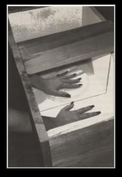
MT is a therapy that uses a mirror to provide a visual illusion to the mind, to trick the brain into thinking that the reflection seen is the patient’s limb doing the actions and if the mind believes this then these cortical changes can occur. MT not only exploits the phenomenon of tricking the brain, but patients must observe their actions; it also activates the mirror neuron system where vicarious mimicry can cause brain reorganisation [4]. Many patients suffering from stroke have little or no movement in their affected limb with over three-quarters of stroke survivors reporting arm weakness [14]. Attempting to rehabilitate this limb is very difficult as they either have insufficient movement in the limb required for rehabilitation or cannot move it at all, so it is not possible to rehab. MT is a type of intervention used in stroke rehabilitation (Fig.) originally developed for Phantom Limb Pain (a Mirror Box is often referred to as a ‘VR box’) [4]. To understand the purpose of the mirror box intervention, it is worth looking at the exact set-up of the original mirror box [4]: “Our ‘virtual reality box’ was constructed by simply placing a 2” by 2” mirror vertically inside the middle of a cardboard box, so that it was perpendicular to the patient’s chest and its upper end was almost touching the chin. The top and face of the box were removed to afford the patient full view of the reflection of his normal hand in the mirror. For patients with a shoulder level disarticulation, a much taller mirror was used.” With an understanding with the setup this allows for better context on the implementation on why the patient in MT performs specific actions: The patient moves the unaffected limb. The patient views the reflection of the unaffected limb in the mirror. A visual illusion is created of the affected limb. Positive feedback for the motor cortex [83].
Before the first session, patients should have enough instructions about the background and aims of MT as well as possible side effects of the treatment. The MT protocol states patients should be able to engage in this kind of treatment and understand that they will imagine that the mirror image is their affected limb. There are indications that the intensity or vividness of the “mirror illusion” may predict the outcomes of the treatment [35]. Identifiers or distractions which present a vivid image such as jewellery and other visual marks like scars, tattoos or birthmarks should be removed or covered to make it easier for the patient to perceive the reflection as their affected limb when looking into the mirror. To create a realistic mirror illusion, place the injured limb behind the mirror in a secure and preferably comfortable posture. For example, the non-affected limb should be positioned in a similar position as the affected limb, as this facilitates the intensity of the mirror illusion. The environment should be free of other stimuli that attract the patients’ attention. For the same reason, at least the first sessions should be delivered individually instead of in a group [35]. The mirror should be big enough to cover the entire affected limb. When choosing a mirror, it should provide a coherent mirror image without notable distortion. Generally, the mirror is positioned in front of the patient’s midline so that the mirror fully covers the affected limb, and the reflection of the unaffected limb is easily seen. Aside from the items needed for functional motor training, exercise materials with additional sensory input might be employed, particularly in individuals with impaired body perception. [35].
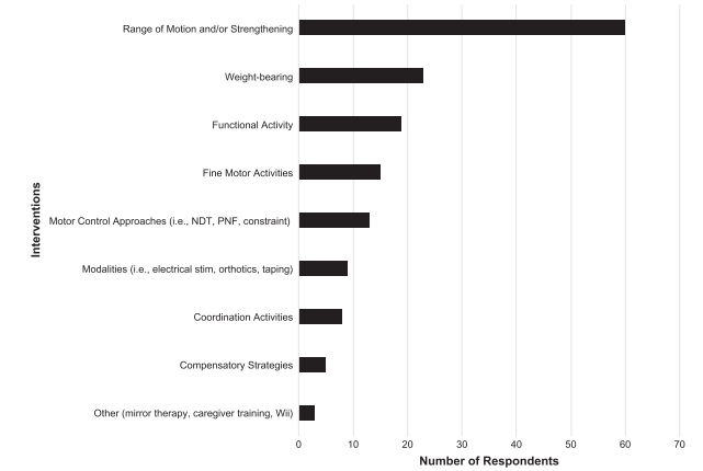
Results suggest that observing one hand in the mirror is easier to imagine movements by motor imagery in comparison to no mirror. Also, a great increase in MEP amplitudes occurred when the movement was assessed even though background EMG activities were absent. These findings point to some probable neural mechanisms [84]. The mirror neuron system could provide an additional or alternative source of information to motor training that may be useful to promote recovery from stroke. Current work will further inform clinical applications in rehabilitation with more evidence supporting methods based on mirror neurons for stroke recovery [85]. MT was the least frequently used intervention used in the survey compared to other types of intervention [86] (Appendix G). Seventy-three occupational therapy practitioners completed the survey. When asked about interventions less than 5 responded with “Other (mirror therapy, caregiver training, Wii)”. The three highest ranked “reported strategies” were “Patient’s interest and goals”, “Education in home program” and “Incorporation of functional activities”.
A recent systematic review of MT for improving motor function after stroke included 62 studies with a total of 1,982 participants that compared MT with other interventions [87]. Its objective was to summarise the effectiveness of MT compared with no treatment, placebo or sham therapy, or other treatments for improving motor function and motor impairment after stroke. It aimed to assess the effects of MT on activities of daily living, pain, and visuospatial neglect. Results of the review indicate that there is moderate evidence for the effectiveness of MT for people after stroke in terms of improving motor function and motor impairment of the upper and lower extremity, as well as improving activities of daily living. When MT is compared to sham (analogous to placebo) interventions, the effects on motor function were more prominent. MT could be applied as an additional intervention in the rehabilitation of people after stroke. There is no definite conclusion on efficacy if MT replaced other interventions for improving motor function of the arm or leg, or both [87].
The review’s implications for research suggest a positive effect of MT after stroke but suffer from methodological problems such as their small sample sizes and lack of proper reporting of relevant factors for research. As such, there is an urgent need for “Further research is needed, with larger methodologically-sound studies.” [87]. These studies should not deliver MT as an adjunct but should compare it to other routinely applied therapies. Further research should also address specific questions about the optimal dose, frequency, and duration of MT [87]. It is suggested that MT is more effective for stroke patients with severe paresis or even flaccid upper limb [5]; other studies and clinical experience suggest patients with better motor ability also benefit from treatment [87], [88]. Eligible patients should possess the cognitive and verbal abilities to focus for ten minutes on the mirror reflection and follow the instruction given by the therapist. For those with vision impairments, therapists should determine if a patient can see a clear image of the entire limb in the mirror. Patients with visuospatial neglect should be able to turn their head toward the mirror image when asked and keep their attention focused on the mirrored image for five to ten minutes. Patients should have enough trunk control and be able to sit unsupervised for the duration of the treatment. Patients with cardiopulmonary abnormalities, who are not able to sit for the duration of the therapy, are not eligible for this kind of treatment. The non-affected limb should ideally have a reasonable and pain-free range of motion. Severe constraints of the non-affected limb could hamper the execution of MT exercises [35].
The main reason to develop a practice-based protocol for MT stemmed from its inconsistency when used in clinical situations as stated by Genius et al. in the “Practical protocol for stroke rehabilitation” [35]. The present MT protocol, and the system MINERVA, viewed as a framework, is not a predefined recipe for all. The framework, however, leaves enough room for the therapist to adjust the protocol and tailor it to the abilities and preferences of the patient. This way, the clinical experience and the preferences of therapists are incorporated into the protocol as well, making it easier to use the protocol in everyday practice. A critical mind is still required [35].
Existing evidence [15], [87], [88] supports the positive effects of MT in stroke patients in the following domains: improving motor function and ADLs, reducing pain, reducing neglect, and reducing sensory impairment. The effects of MT on spasticity have not been established in clinical studies, although clinical experience suggests it may help with the short-term reduction of spasticity in patients with stroke [35]. The available literature [87], [88] recommends performing MT at least once daily with a minimum duration of ten minutes. The maximum duration of each session is dependent on the cognitive abilities of the individual patient and any adverse side effects, but in most cases, will be around 30 minutes. However, a 2020 pilot by Hsieh et al. [84] reports MT as the least effective of the approaches studied and reports preliminary findings favouring action observation therapy [89]. Imagination, i.e., copying someone else’s actions even as a thought, can evoke the mirror neuron system through its interaction with neural motor areas, which lends itself well to MT. Combined with other therapies and strategies, MT has shown promising results for stroke rehabilitation. The interaction with vision, proprioception and motor command for voluntary movement, promotes mirror neuron excitability and cortical rearrangement, resulting in functional movement. Despite the potential observed for MT by a 2013 study led by Carvalho [86], many areas of the brain are not activated in MT and could be compromised. Patients experience fatigue and attention level decrease, which can cause a lack of concentration when executing MT tasks [90]. To understand the cortical mechanisms of MT after stroke [91]: “First, measure cortical activity directly during MT using MEG and to examine the differential effects in controls and stroke patients. Our results suggest a rebalancing of MRBD between hemispheres in stroke patients. Alterations in beta oscillations have links to changes in intracortical GABAergic inhibitory function, and it is interesting to speculate on whether our results reflect an MT-enhanced potential for experience-dependent plasticity within motor networks. In future studies, it would be of interest to determine whether the effects of MT on cortical physiology that we have observed here are necessary (i.e., biomarkers) for a beneficial effect on motor function.” The design should consider these factors and work around them. A possible implementation to counter a lack of focus would be a shorter, sharper and more engaging experience for the patient to reduce the extraneous cognitive load from session instructions. The system should be designed to take into consideration that a patient may experience fatigue during a session and what the correct response should be.
Adding haptic sensations can also trick the brain, as in the rubber hand illusion [92]. The rubber hand illusion works by the patient visually observing a rubber hand being stroked by a paint brush while simultaneously the patient’s hand, which is out of view, is being stroked by a paint brush in the exact same area and time. The purpose of this simultaneous stroking on both the real and rubber hand is to create haptic sensory congruence and since the eyes can only see the rubber hand, an association occurs that the rubber hand is the patient’s hand. The right parietal lobe is a critical component of the brain’s navigation system. It is a flexible boundary of self and other objects; like how a driver can ‘feel’ the road through the pedals[93]. Merely making a digital version of MT, which already exists in reality, and which has been shown to work [94],[95],[96], will not maximise the potential of VR. According to Park et al. [97], when the sub items of the capacity to perform daily living tasks in the mirror group were compared to those in the control group, statistically significant changes in the area of self-care occurred.
Robotics can be useful, but it is outside the scope of MINERVA’s design. Nam et al. [98] mention that it may be useful for supratentorial (region of the brain) stroke patients to facilitate recovery of proprioceptive (sense of location) deficit and hemineglect symptoms. Kim et al. [99] state, “It has a disadvantage of shifting weight and leaning towards the unaffected side during therapy.” The current system developed overcomes this issue as the mirror plane is always relative to the patient, no other systems developed can do this currently. This implementation prevents distortion by any displacement from the patient and the mirror plane’s optimal position. Since the mirroring plane can move with the patient this creates opportunities which cannot be done in traditional mirroring methods, and this is the first VRMT system to attempt it.
Summary
Stroke rehabilitation works and there are a variety of interventions each with their advantages and disadvantages. Neuroplasticity, motor control, and learning mechanisms provide a clearer insight into how rehabilitation works in theory and practice through the literature available. MT was selected as there is promising evidence of its effectiveness for rehabilitation and is likely to be most beneficial for patients who have little to no movement in the impaired limb, which is the primary type of stroke patients that the system intends to rehabilitate.
References (65)
- [4] V. S. Ramachandran and D. Rogers-Ramachandran, “Synaesthesia in Phantom Limbs Induced with Mirrors,” Proc. R. Soc. B Biol. Sci., vol. 263, no. 1369, pp. 377–386, 1996, doi: 10.1098/rspb.1996.0058. ↩
- [6] L. Non-use, “Encyclopedia of Clinical Neuropsychology,” Encycl. Clin. Neuropsychol., pp. 2–4, 2018, doi: 10.1007/978-3319-56782-2. ↩
- [15] S. Doyle, K. McKenna, S. Bennette, and S. Fasoli, “Interventions for sensory impairment in the upper limb after stroke (Review),” no. 6, 2010, doi: 10.1002/14651858.CD006331.pub2. ↩
- [16] S. Helpline, “Physiotherapy after stroke,” stroke Assoc., vol. 16, no. 1, pp. 1–8, 2017. ↩
- [25] A. Constans and J. Little, Body by Science: A Research Based Program for Strength Training, Body building, and Complete Fitness in 12 Minutes a Week: A Research Based Program to Get the Results You Want in 12 Minutes a Week, vol. 17, no. 19. McGraw-Hill 2012, [Online]. Available: Education, 2009. ↩
- [28] N. S. Ward, F. Brander, and K. Kelly, “Intensive upper limb neurorehabilitation in chronic stroke: Outcomes from the Queen Square programme,” J. Neurol. Neurosurg. Psychiatry, vol. 90, no. 5, pp. 498–506, 2019, doi: 10.1136/jnnp-2018319954. ↩
- [35] J. Genius, S. Roß, S. Uhr, S. Braun, A. Rothgangel, and P. Verlag, “Practical protocol for stroke rehabilitation,” Pain Pract., vol. 16, no. 4, pp. 422–434, 2013, doi: 10.12855/ar.sb.mirrortherapy.e2013. ↩
- [42] H. Johansen-berg, “Reorganisation and modulation of the human sensorimotor system: implications for recovery of motor function after stroke,” Philosophy, pp. 1–35, 2001, doi: 10.1589/jpts.28.1403. ↩
- [43] R. L. Harvey, “Improving poststroke recovery: Neuroplasticity and task-oriented training,” Curr. Treat. Options Cardiovasc. Med., vol. 11, no. 3, pp. 251–259, 2009, doi: 10.1007/s11936-009-0026-4. ↩
- [44] Y. J. Kang, J. Ku, H. J. Kim, and H. K. Park, “Facilitation of Corticospinal Excitability According to Motor Imagery and Mirror Therapy in Healthy Subjects and Stroke Patients,” Ann. Rehabil. Med., vol. 35, no. 6, p. 747, 2011, doi: 10.5535/arm.2011.35.6.747. ↩
- [45] E. Cakar et al., “The relationships of motor-evoked potentials to hand dexterity, motor function, and spasticity in chronic stroke patients: a transcranial magnetic stimulation study,” Acta Neurol. Belg., vol. 116, no. 4, pp. 481–487, 2016, doi: 10.1007/s13760-016-0633-2. ↩
- [46] S. Bestmann and J. W. Krakauer, “The uses and interpretations of the motor-evoked potential for understanding behaviour,” Exp. Brain Res., vol. 233, no. 3, pp. 679–689, 2015, doi: 10.1007/s00221-014-4183-7. ↩
- [47] T. H. Murphy and D. Corbett, “Plasticity during stroke recovery: From synapse to behaviour,” Nat. Rev. Neurosci., vol. 10, no. 12, pp. 861–872, 2009, doi: 10.1038/nrn2735. ↩
- [48] Stress: Physiology, Biochemistry, and Pathology, 1 edition. Academic Press, 2019. ↩
- [49] L. Nummenmaa, R. Hari, J. K. Hietanen, and E. Glerean, “Maps of subjective feelings,” Proc. Natl. Acad. Sci. U. S. A., vol. 115, no. 37, pp. 9198–9203, 2018, doi: 10.1073/pnas.1807390115. ↩
- [50] C. Foulon et al., “Advanced lesion symptom mapping analyses and implementation as BCBtoolkit,” Gigascience, vol. 7, no. 3, pp. 1–17, 2018, doi: 10.1093/gigascience/giy004. ↩
- [51] “Learned Non-Use.” https://www.cimt.co.uk/adults/theory-of-cimt/learned-non-use.php. ↩
- [52] J. J. Radley et al., “Repeated stress induces dendritic spine loss in the rat medial prefrontal cortex,” Cereb. Cortex, vol. 16, no. 3, pp. 313–320, 2006, doi: 10.1093/cercor/bhi104. ↩
- [53] O. Hurtado et al., “A chronic treatment with CDP-choline improves functional recovery and increases neuronal plasticity after experimental stroke,” Neurobiol. Dis., vol. 26, no. 1, pp. 105–111, 2007, doi: 10.1016/j.nbd.2006.12.005. ↩
- [54] B. B. Johansson, “Brain Plasticity and Stroke Rehabilitation : The Willis Lecture,” Stroke, vol. 31, no. 1, pp. 223–230, 2000, doi: 10.1161/01.STR.31.1.223. ↩
- [55] R. Schmidt, Motor Control and Learning 6th Edition with Web Resource: A Behavioral Emphasis, 6th ed. Human Kinetics Publishers, 2018. ↩
- [56] P. L. Weiss, E. A. Keshner, and M. F. Levin, Virtual Reality for Physical and Motor Rehabilitation (Virtual Reality Technologies for Health and Clinical Applications), 2014 editi. Springer, 2016. ↩
- [57] S. M. Hatem et al., “Rehabilitation of Motor Function after Stroke: A Multiple Systematic Review Focused on Techniques to Stimulate Upper Extremity Recovery,” Front. Hum. Neurosci., vol. 10, no. September, pp. 1–22, 2016, doi: 10.3389/fnhum.2016.00442. ↩
- [58] S. J. Page and H. Peters, “Mental practice: Applying motor PRACTICE and neuroplasticity principles to increase upper extremity function,” Stroke, vol. 45, no. 11, pp. 3454–3460, 2014, doi: 10.1161/STROKEAHA.114.004313. ↩
- [59] Intercollegiate Stroke Working Party, National clinical guideline for stroke, 5th ed., no. July. 2016. ↩
- [60] NICE, “Stroke rehabilitation in adults,” Natl. Institue Heal. Care Excell., no. April, 2016, doi: 10.17674/19970854.2018.1.156-165. ↩
- [61] K. S. Hayward and S. G. Brauer, “Dose of arm activity training during acute and subacute rehabilitation post stroke: A systematic review of the literature,” Clin. Rehabil., vol. 29, no. 12, pp. 1234–1243, 2015, doi: 10.1177/0269215514565395. ↩
- [62] T. West and J. Bernhardt, “Physical activity in hospitalised stroke patients,” Stroke Res. Treat., vol. 2012, no. October 2010, 2012, doi: 10.1155/2012/813765. ↩
- [63] C. English, J. Bernhardt, and S. Hillier, “Circuit class therapy and 7-day-week therapy increase physiotherapy time, but not patient activity: Early results from the CIRCIT trial,” Stroke, vol. 45, no. 10, pp. 3002–3007, 2014, doi: 10.1161/STROKEAHA.114.006038. ↩
- [64] van W. Vloothuis JDM, Mulder M, Veerbeek JM, Konijnenbelt M, Visser-Meily JMA, Ket JCF, Kwakkel G, “CaregiverMediated Exercises for Improving Outcomes After Stroke (Cochrane Review) [With Consumer Summary],” Cochrane Database Syst. Rev., vol. 12, no. 12, pp. 1–117, 2016, doi: 10.1002/14651858.CD011058.pub2.www.cochranelibrary.com. ↩
- [65] G. Kwakkel, “Intensity of practice after stroke: More is better,” Schweizer Arch. fur Neurol. und Psychiatr., vol. 160, no. 7, pp. 295–298, 2009, doi: 10.1080/09638280500534861. ↩
- [66] C. Han, Q. Wang, P. Meng, and M. Qi, “Effects of intensity of arm training on hemiplegic upper extremity motor recovery in stroke patients: a randomized controlled trial,” Clin. Rehabil., vol. 27, no. 1, pp. 75–81, 2013, doi: 10.1177/0269215512447223. ↩
- [67] J. Langan, H. Subryan, I. Nwogu, and L. Cavuoto, “Reported use of technology in stroke rehabilitation by physical and occupational therapists,” Disabil. Rehabil. Assist. Technol., vol. 0, no. 0, pp. 1–7, 2017, doi: 10.1080/17483107.2017.1362043. ↩
- [68] R. Teasell et al., “Canadian Stroke Best Practice Recommendations: Rehabilitation, Recovery and Community Participation following Stroke. Part One: Rehabilitation and Recovery following Stroke; 6 th Edition Update 2019.e,” Int. J. Stroke, vol. 0, no. 0, pp. 1–26, 2019, doi: 10.1177/1747493019897843. ↩
- [69] A. Pollock et al., “Interventions for improving upper limb function after stroke (Review),” Cochrane Database Syst. Rev., vol. 11, no. 11, pp. CD010820–CD010820, 2014, doi: 10.1002/14651858.CD010820.pub2.www.cochranelibrary.com. ↩
- [70] G. A. Eskes et al., “Canadian Stroke Best Practice Recommendations: Mood, Cognition and Fatigue Following Stroke practice guidelines, update 2015,” Int. J. Stroke, vol. 10, no. 7, pp. 1130–1140, 2015, doi: 10.1111/ijs.12557. ↩
- [71] S. H. Lin and T. P. Dionne, “Interventions to Improve Movement and Functional Outcomes in Adult Stroke Rehabilitation: Review and Evidence Summary,” J. Particip. Med., vol. 10, no. 1, p. e3, 2018, doi: 10.2196/jopm.8929. ↩
- [72] K. Kelly and F. Brander, “An Integrated Therapeutic Approach to Intensive Upper Limb Rehabilitation,” 2018, no. June. ↩
- [73] National Clinical Guideline Centre, “Stroke Rehabilitation - Long term rehabilaition after stroke,” NICE Guid., vol. Clinical g, 2013, [Online]. Available: http://www.nice.org.uk/nicemedia/live/14182/64094/64094.pdf. ↩
- [74] N. Ward and Q. Square, “Intensive upper limb neurorehabilitation in chronic stroke: outcomes from the Queen Square programme,” Neurol Neurosurg Psychiatry, pp. 498–506, 2019, doi: http:// dx. doi. org/ 10. 1136/ jnnp- 2018- 319954. ↩
- [75] R. Teasell, J. Bitensky, K. Salter, and N. A. Bayona, “The role of timing and intensity of rehabilitation therapies,” Top. Stroke Rehabil., vol. 12, no. 3, pp. 46–57, 2005, doi: 10.1310/ETDP-6DR4-D617-VMVF. ↩
- [76] N. G. Schoepp et al., “Dose-response of task-specific upper limb training in people at least 6 months post stroke: A Phase II, single-blind, randomized, controlled trial,” vol. 55, no. 33, pp. 9557–9561, 2017, doi: 10.1002/anie.201602763.Digital. ↩
- [77] B. French et al., “Repetitive task training for improving functional ability after stroke (Review),” Cochrane Database Syst. Rev, no. 11, 2016, doi: 10.1002/14651858.CD006073.pub3.www.cochranelibrary.com. ↩
- [78] L. A. Connell, N. E. McMahon, J. J. Eng, and C. L. Watkins, “Prescribing upper limb exercises after stroke: A survey of current UK therapy practice,” J. Rehabil. Med., vol. 46, no. 3, pp. 212–218, 2014, doi: 10.2340/16501977-1268. ↩
- [79] R. N. Carpinelli and R. M. Otto, “Strength training. Single versus multiple sets,” Sport. Med., vol. 26, no. 2, pp. 73–84, 1998, doi: 10.2165/00007256-199826020-00002. ↩
- [80] J. Crozier et al., “High-Intensity Interval Training After Stroke: An Opportunity to Promote Functional Recovery, Cardiovascular Health, and Neuroplasticity,” Neurorehabil. Neural Repair, vol. 32, no. 6–7, pp. 543–556, 2018, doi: 10.1177/1545968318766663. ↩
- [81] S. A. Boudreau, D. Farina, and D. Falla, “The role of motor learning and neuroplasticity in designing rehabilitation approaches for musculoskeletal pain disorders,” Man. Ther., vol. 15, no. 5, pp. 410–414, 2010, doi: 10.1016/j.math.2010.05.008. ↩
- [82] K. Hutchins, “Exercise vs Recreation,” content/uploads/2010/11/Exercise-vs-Recreation.pdf. ↩
- [83] A. Cacchio, E. De Blasis, V. De Blasis, V. Santilli, and G. Spacca, “Mirror therapy in complex regional pain syndrome type 1 of the upper limb in stroke patients.,” Neurorehabilitation & Neural Repair, vol. 23, no. 8, pp. 792-799 8p, 2009, doi: 10.1177/1545968309335977. ↩
- [84] K. Fukumura, K. Sugawara, S. Tanabe, J. Ushiba, and Y. Tomita, “Influence of mirror therapy on human motor cortex,” Int. J. Neurosci., vol. 117, no. 7, pp. 1039–1048, 2007, doi: 10.1080/00207450600936841. ↩
- [85] K. A. Garrison, C. J. Winstein, and L. Aziz-Zadeh, “The mirror neuron system: A neural substrate for methods in stroke rehabilitation,” Neurorehabil. Neural Repair, vol. 24, no. 5, pp. 404–412, 2010, doi: 10.1177/1545968309354536. ↩
- [86] E. V. D. Brown et al., “Home programs for upper extremity recovery post-stroke: A survey of occupational therapy practitioners,” Top. Stroke Rehabil., vol. 9357, no. 8, pp. 573–578, 2017, doi: 10.1080/10749357.2017.1366013. ↩
- [87] H. Thieme et al., “Mirror therapy for improving motor function after stroke ( Review ),” Cochrane Database Syst. Rev., no. 7, 2018, doi: 10.1002/14651858.CD008449.pub3.www.cochranelibrary.com. ↩
- [88] A. S. Rothgangel, S. M. Braun, A. J. Beurskens, R. J. Seitz, and D. T. Wade, “The clinical aspects of mirror therapy in rehabilitation: A systematic review of the literature,” Int. J. Rehabil. Res., vol. 34, no. 1, pp. 1–13, 2011, doi: 10.1097/MRR.0b013e3283441e98. ↩
- [89] Y. W. Hsieh, Y. H. Lin, J. D. Zhu, C. Y. Wu, Y. P. Lin, and C. C. Chen, “Treatment Effects of Upper Limb Action Observation Therapy and Mirror Therapy on Rehabilitation Outcomes after Subacute Stroke: A Pilot Study,” Behav. Neurol., vol. 2020, 2020, doi: 10.1155/2020/6250524. ↩
- [90] D. Carvalho et al., “The mirror neuron system in post-stroke rehabilitation,” Int. Arch. Med., vol. 6, no. 1, pp. 1–7, 2013, doi: 10.1186/1755-7682-6-41. ↩
- [91] H. E. Rossiter, M. R. Borrelli, R. J. Borchert, D. Bradbury, and N. S. Ward, “Cortical mechanisms of mirror therapy after stroke,” Neurorehabil. Neural Repair, vol. 29, no. 5, pp. 444–452, 2015, doi: 10.1177/1545968314554622. ↩
- [92] M. Botvinick and J. Cohen, “Rubber hands ‘feel’ touch that eyes see.,” vol. 391, no. February, p. 1998, 1998, doi: 10.1038/35784. ↩
- [93] S. Kotler and J. Wheal, Stealing Fire: How Silicon Valley, the Navy SEALs, and Maverick Scientists Are Revolutionizing the Way We Live and Work, Reprint ed. Dey Street Books, 2018. ↩
- [94] H. T. Regenbrecht, E. A. Franz, G. McGregor, B. G. Dixon, and S. Hoermann, “Beyond the looking glass: Fooling the brain with the augmented mirror box,” Presence Teleoperators Virtual Environ., vol. 20, no. 6, pp. 559–576, 2011, doi: 10.1162/PRES_a_00082. ↩
- [95] K. Hartman and E. L. Altschuler, “Mirror Therapy for Hemiparesis Following Stroke: A Review,” Curr. Phys. Med. Rehabil. Reports, vol. 4, no. 4, pp. 237–248, 2016, doi: 10.1007/s40141-016-0131-8. ↩
- [96] T. In, K. Lee, and C. Song, “Virtual Reality Reflection Therapy Improves Balance and Gait in Patients with Chronic Stroke: Randomized Controlled Trials,” Med. Sci. Monit., vol. 22, pp. 4046–4053, 2016, doi: 10.12659/MSM.898157. ↩
- [97] J.-Y. Park, M. Chang, K.-M. Kim, and H.-J. Kim, “The effect of mirror therapy on upper-extremity function and activities 2009, [Online]. Available: http://ren-ex.com/wp- of daily living in stroke patients,” J. Phys. Ther. Sci., vol. 27, no. 6, pp. 1681–1683, 2015, doi: 10.1589/jpts.27.1681. ↩
- [98] H. S. Nam et al., “Recovery of Proprioception in the Upper Extremity by Robotic Mirror Therapy: a Clinical Pilot Study for Proof of Concept,” @BULLET J Korean Med Sci, vol. 32, pp. 1568–1575, 2017, doi: 10.3346/jkms.2017.32.10.1568. ↩
- [99] J. Kim, J. Yi, and C.-H. Song, “Kinematic analysis of head, trunk, and pelvic motion during mirror therapy for stroke patients,” J. Phys. Ther. Sci., vol. 29, no. 10, pp. 1793–1799, 2017, doi: 10.1589/jpts.29.1793. ↩
Appendices (5)
Appendix C. Canadian Stroke Best Practice evidence rating ↩
Canadian Stroke Best Practice Therapy evidence rating
| Rating | Description |
|---|---|
| A | Evidence from a meta-analysis of randomized controlled trials or consistent findings from two or more randomized controlled trials, where it is obvious the desirable effects outweigh undesirable effects or undesirable effects outweigh desirable effects |
| B | Evidence from a single randomized controlled trial or consistent findings from two or more well- designed non- randomized and/or non-controlled trials, and large observational studies. Desirable effects outweigh or are closely balanced with undesirable effects or undesirable effects outweigh or are closely balanced with desirable effects. |
| C | Writing group consensus and/or supported by limited research evidence. Desirable effects outweigh or are closely balanced with undesirable effects or undesirable effects outweigh or are closely balanced with desirable effects, as determined by writing group consensus. Recommendations assigned a Level- C evidence may be key system drivers supporting other recommendations, and some may be expert opinion based on common, new, or emerging evidence or practice patterns. |
Canadian Stroke Best Practice Therapy evidence level.
| # | Specfic Therapy | Evidence Level |
|---|---|---|
| i | ROM exercises (passive and active-assisted) that includes placement of the upper limb in a variety of appropriate and safe positions within the patient’s visual field should be provided. Refer to Recommendation ‘‘Recommendations on management of shoulder pain and complex regional pain syndrome (CRPS) following stroke’’ for additional information. |
Level C |
| ii | Following assessment to determine if they are suitable candidates, patients should be encouraged to engage in mental imagery to enhance upper-limb, sensorimotor recovery. |
Early-Level A; Late-Level B |
| iii | Functional Electrical Stimulation (FES) targeted at the wrist and forearm muscles should be considered to reduce motor impairment and improve function. |
Early-Level A; Late-Level A |
| iv | Traditional or modified constraint-induced movement therapy should require consideration for a select group of patients who demonstrate at least 200 of active wrist extension and 100 of active finger extension, with minimal sensory deficits and normal cognition. |
Early-Level A; Late-Level A |
| v | Mirror therapy requires consideration as an adjunct to motor therapy for patients with very severe paresis. It may help to improve upper extremity motor function and ADLs. |
Early-Level A; Late-Level A |
| vi | Despite mixed evidence, sensory stimulation (e.g. transcutaneous electrical nerve stimulation, acupuncture, biofeedback) can be considered as an adjunct to improve upper extremity function. |
Evidence Level B |
| vii | Virtual reality, including both immersive technologies such as head-mounted or robotic interfaces and non- immersive technologies such as gaming devices can be used as adjunct tools to other rehabilitation therapies as a means to provide additional opportunities for engagement, feedback, repetition, intensity, and task- oriented training. |
Evidence Level: Early-Level A; Late-Level A |
| viii | Therapists should consider supplementary training programs aimed at increasing the active movement and functional use of the affected arm between therapy sessions, e.g. Graded Repetitive Arm Supplementary Program suitable for use during hospitalization and at home. |
Evidence Level: Early-Level B; Late-Level C |
| ix | Strength training requires consideration for persons with mild to moderate upper extremity impairment for improvement in grip strength. Strength training does not aggravate tone or pain. |
Grip strength; Early-Level A; Late-Level A, Strength training; Level A |
| x | Bilateral arm training is not recommended over unilateral arm training to improve upper extremity motor function. |
Level A |
| xi | Non-invasive brain stimulation, including repetitive transcranial magnetic stimulation (rTMS) and transcranial direct current stimulation (tDCS) could be considered as an adjunct to upper extremity therapy. |
Level A (rTMS); Level B (tDCS) |
| xii | For patients who are unable to produce any voluntary muscle activity in the affected upper limb, the patient (and caregiver) should be taught compensatory techniques and be provided with adaptive equipment to enable basic ADLs. It is reasonable to continue teaching compensatory techniques until the patient can manage basic ADLs independently or until recovery of active movement occurs. |
Adaptive equipment to enable basic ADLs (Evidence Level B). until recovery of active movement occurs (Evidence Level C). |
Appendix D. Intervention, grade of evidence for Outcome ↩
Appendix E. Queens Square Upper Limb Fugl-Meyer Results ↩
Appendix F. Queen Square UL Summary ↩
Appendix G. Interventions reported as used in upper extremity home programs ↩